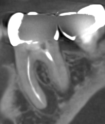
This image shows a cross-section of a CBCT image showing an infected lower right first molar that was previously treated with a root canal. The large black halo on the front root (left) shows an infected abscess in the patient’s jawbone.
Our office is highly experienced in using the latest dental imaging to evaluate root canals and identify the presence of a recurrent infection. Many times the patient has no localized pain or discomfort in the tooth. The CBCT equipment we have in our office is vital to identifying infections which can cause a systemic health concern even if the infection is without localized pain.
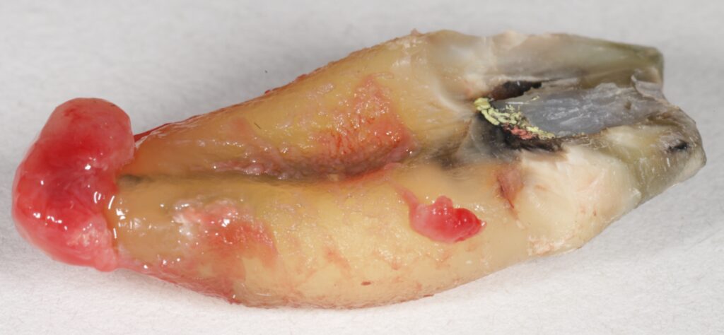
This image shows a piece of the same tooth illustrated above that was surgically removed. The large red ball of tissue at the end of the root (left) is the infected abscess that was surgically removed along with the bad tooth.