In-Office CBCT Imaging and Evaluation

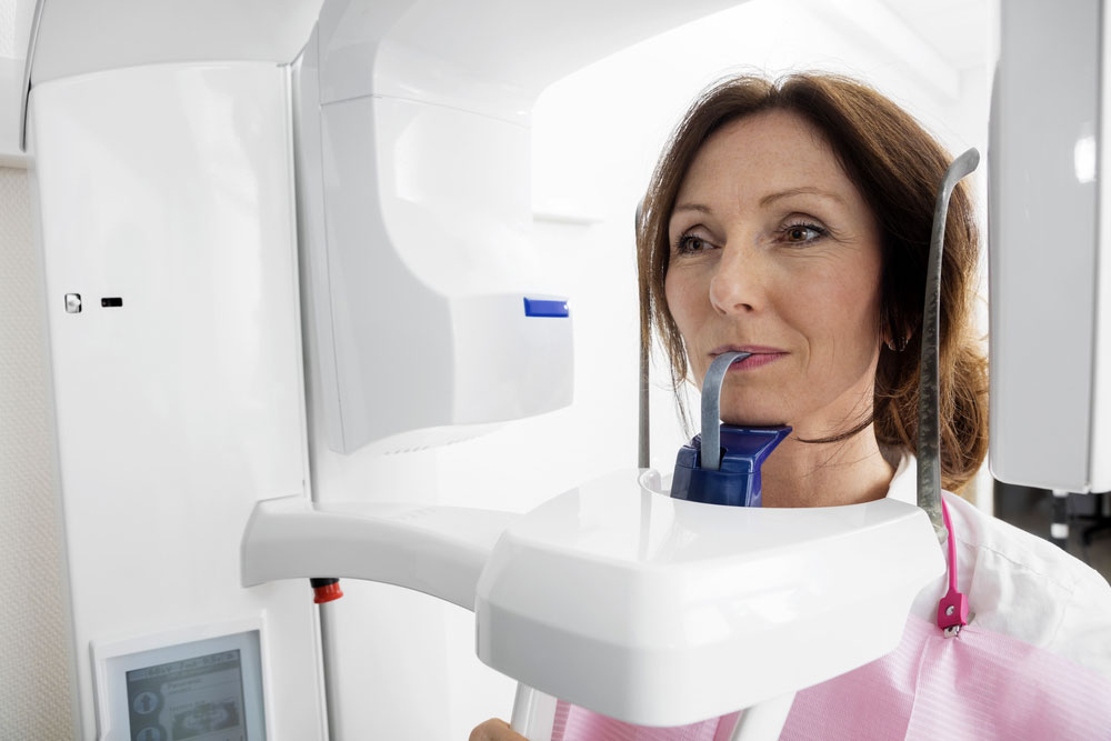
Dr. Tatarin practices dentistry with a progressive approach and for that reason our office utilizes the latest in dental imaging. Our office has the latest Planmeca Cone Beam Computed Tomography (CBCT) to best identify dental concerns that the patient may be unaware of that may have an adverse effect on a patient’s overall health. Our CBCT imaging technology is incredibly valuable to our ability to correctly identify dental concerns including:

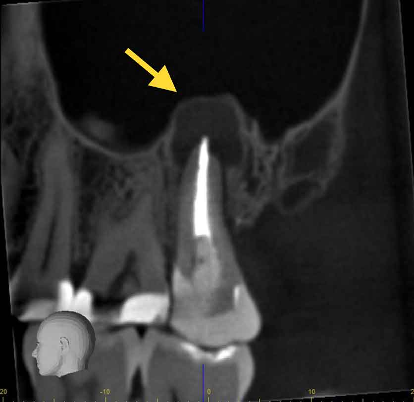
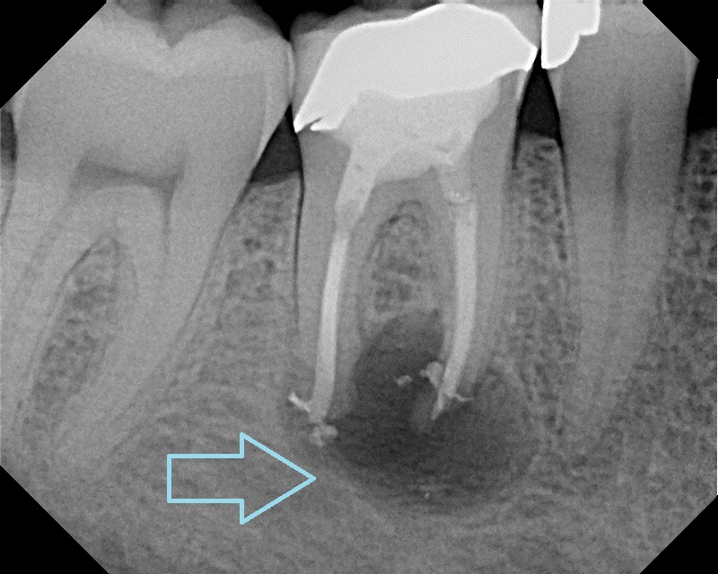
  • Identify infected root canal treated teeth or other necrotic teeth that may be harboring harmful bacteria that can increase the body’s inflammatory markers.
  • Identify the true 3-dimensional extent of dental infections to guarantee their complete removal
  • Identify the extent of dental caries or other pathology
  • Evaluate 3-dimensional tooth morphology that is missed on conventional 2-dimensional x-rays.
  • Evaluate bone density and bone levels as they relate to periodontal disease
  • Evaluate 3-dimensional bone contours and anatomy for safe and successful dental implant procedures.
CBCT Imaging
This image shows our state-of-the-art Planmeca CBCT in action. Taking a CBCT image on a patient is very easy and takes just a few seconds. The patient sits on a stool and rests their head against the machine. The CBCT spins arounds the patients head to capture a 3-dimensional image.

Below are just a few examples of clinical cases we have identified and treated in our office using CBCT imaging.

Infected tooth found using CBCT Imaging
This image depicts an infected root-canal treated tooth visualized on our CBCT. The black halo visualized at the apex (root tip) of the tooth is filled with bacteria, pus, and infected tissue that is surgically removed during extraction. The extent of this pathological lesion could potentially be missed without advanced CBCT imaging.
Infected Molar Found Furing CBCT Imaging and Evaluation
This CBCT image shows a re-infected lower molar after root-canal treatment. In this example, excessive sealer extruded out of the root during the root canal procedure which contributed to the colonization of bacteria. The large black halo is infected bone.

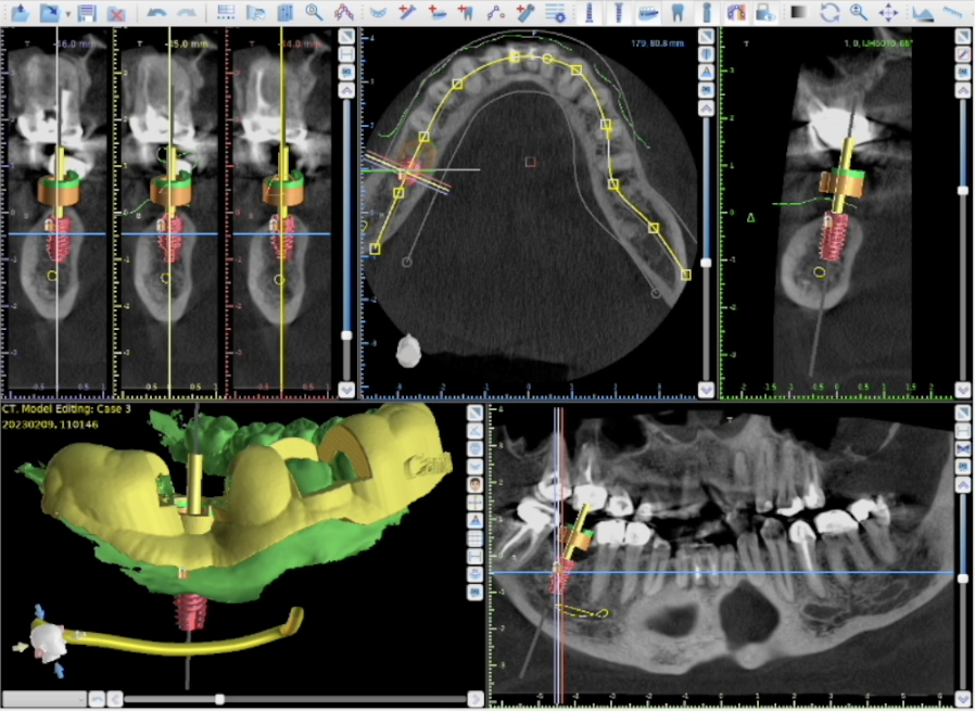
We also use our 3-D CBCT technology to digitally plan surgical dental implant placement

This image shows a CBCT image loaded in to our digital implant planning software. With this technology we can guarantee safe and ideal implant placement for the best long term implant success because we can visualize the exact 3-dimensional anatomy before the procedure.